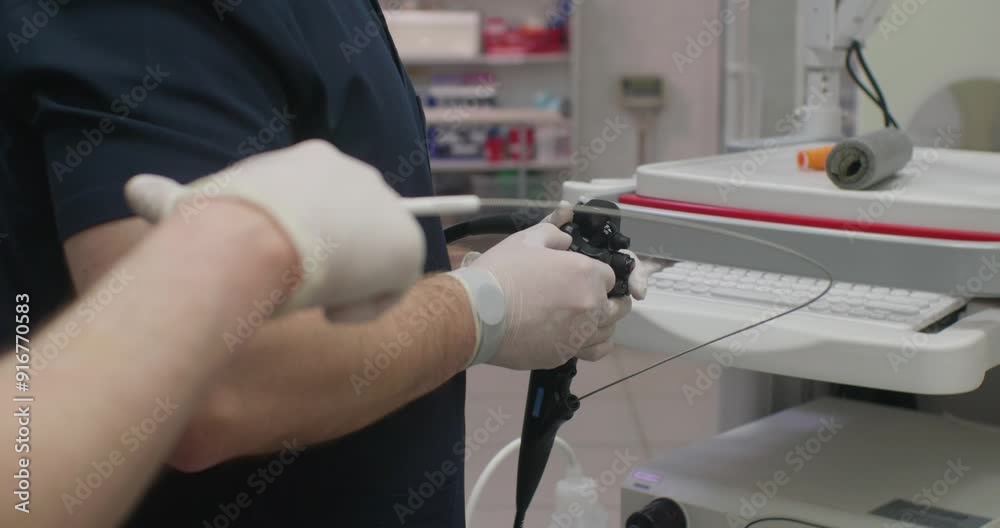
**Reason Behind the Horrifying Images Captured During a Routine Endoscopic Exam** For many Americans, the idea of a routine medical procedure might feel routine in name only—yet when vivid images surface online, they spark silence, curiosity, and cautious concern. One of the most discussed phenomena today is the “horrifying images captured during a routine endoscopic exam.” Though medical image visibility may seem alarming, understanding what causes these visuals helps demystify a procedure that plays a crucial role in diagnostic health care. This article explores why these striking images appear, their medical significance, and what patients and readers should know—without speculation, clickbait, or sensationalism. ### Why the Trend Is Gaining Attention in the US In recent years, digital transparency around healthcare has grown alongside increased patient engagement and online medical forums. The phrase “horrifying images captured during a routine endoscopic exam” now regularly surfaces in search queries, reflecting a mix of genuine concern and the viral nature of unusual findings in medical imaging. This visibility is amplified by social media’s role in spreading medical anecdotes, often without clinical context. While attention often focuses on the striking visuals, the deeper reason stems from how these images relate to the interior health of the digestive tract—information increasingly visible to curious patients and those navigating unfamiliar procedures. ### How These Images Actually Happen During an Endoscopy
### Common Questions About the Visible Images **Q: Are these images normal during an endoscopy?** Yes—visible anatomical details like lining patterns or mild mucus are standard and highly informative. **Q: Do these images mean something concerning is happening?** Not by themselves. They often reflect normal structures, but persistent or unusual findings are documented separately during diagnostic review. **Q: Can the images be taken out of context?** Yes. Social media often highlights striking visuals without explaining medical context, contributing to fear or misunderstanding. **Q: Is nothing abnormal behind today’s photos?** No. Medical teams monitor and interpret every visual cue carefully. Isolated images rarely indicate pathology unless followed by specific symptoms. ### Opportunities and Realistic Considerations Understanding why these images surface offers patients confidence and clarity. They reveal a minimally invasive tool used daily—often critical for detecting early abnormalities. For non-experts, the surprise stems from exposure to a side of medicine rarely seen outside clinics. For healthcare providers, these visuals support transparency and education, especially when explaining endoscopy results. While rare, instances of unexpected visual prominence should always prompt follow-up care—not alarm. Medical professionals emphasize context: images alone mean little without clinical correlation. ### What People Commonly Misunderstand A major misconception is that bright or unusual images immediately signal disease. In reality, anatomy varies widely; what seems “horrifying” to a layperson is often typical. Another myth is that endoscopic photos are routinely shared publicly without consent—actual protocols strictly protect privacy and require explicit patient communication. Confusing normal findings with pathology fuels unnecessary anxiety. Clarifying medical terminology, explaining imaging roles, and promoting open provider-patient dialogue reduces fear and builds trust. ### Who This Topic Matters For Understanding this phenomenon touches diverse groups: patients preparing for exams seeking clarity, caregivers researching symptoms, and health professionals guiding conversations. Parents may worry after photos circulate. Anyone questioning diagnostic results needs honest answers. Patients from diverse backgrounds benefit from transparent, accessible information that respects cultural sensitivities and avoids triggering distress. ### A Soft CTA Encouraging Informed Awareness If curiosity leads you further, seek information from trusted medical sources—your gastroenterologist, reputable health websites, or patient education materials. Informed understanding safeguards peace of mind, encouraging proactive but measured health habits. Stay curious, stay informed, and prioritize professional guidance over online speculation. ### Conclusion The “horrifying images” captured during routine endoscopic exams reflect the complex visual landscape of modern diagnostic medicine—not warnings, but windows into internal health. Grounded in physiology and clinical practice, these visuals reinforce the value of early detection and transparency. By understanding their origin and meaning, patients gain clarity and confidence. In an era of instant information, critical thinking and trust in healthcare providers remain the best tools for navigating these moments with calm and awareness.
Understanding this phenomenon touches diverse groups: patients preparing for exams seeking clarity, caregivers researching symptoms, and health professionals guiding conversations. Parents may worry after photos circulate. Anyone questioning diagnostic results needs honest answers. Patients from diverse backgrounds benefit from transparent, accessible information that respects cultural sensitivities and avoids triggering distress. ### A Soft CTA Encouraging Informed Awareness If curiosity leads you further, seek information from trusted medical sources—your gastroenterologist, reputable health websites, or patient education materials. Informed understanding safeguards peace of mind, encouraging proactive but measured health habits. Stay curious, stay informed, and prioritize professional guidance over online speculation. ### Conclusion The “horrifying images” captured during routine endoscopic exams reflect the complex visual landscape of modern diagnostic medicine—not warnings, but windows into internal health. Grounded in physiology and clinical practice, these visuals reinforce the value of early detection and transparency. By understanding their origin and meaning, patients gain clarity and confidence. In an era of instant information, critical thinking and trust in healthcare providers remain the best tools for navigating these moments with calm and awareness. --- *Keyword:** Reason behind the horrifying images captured during a routine endoscopic exam
--- *Keyword:** Reason behind the horrifying images captured during a routine endoscopic exam
Zum Abominable Truth Hidden in Every Silence
The Untold Story of Yakima’s Herald That Shook the Pacific Northwest
This Accidental Wrong Turn Movie got Dangerously Wrong—and You’ll Laugh Out Loud
